| Prev | ICM User's Guide 17.5 ICM X-Ray AutoFit - Automated Model Building into Density | Next |
The ICM X-Ray AutoFit is an automated method to fit a ligand into electron density. The tool combines the powerful ICM docking algorithm with an electron density fitting function.
The input for ICM X-Ray AutoFit is an electron density map in CCP4 format, the protein recpeptor and ligand which can either be drawn or imported into ICM.
Theory
The ICM X-Ray AutoFit method includes the following features:
- Soft docking energy function.
- Intra and inter ligand interaction energy function.
- Weighted electron density contributions.
- The electron density for the fit function is filtered to exclude areas occcupied by the protein receptor atoms.
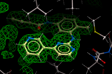
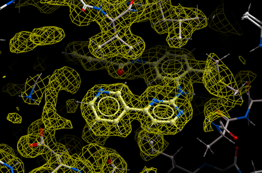
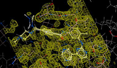
| NOTE: Density from the receptor is automatically filtered out from the analysis. In the picture shown above the green map represents attractive potential. |
The method generates multiple hits for each ligand with a score assigned. It has been demonstrated that improved ligand receptor interactions can be determined by the ICM X-Ray AutoFit method compared to published crystal structures.
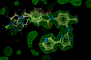
In the figure shown below the interaction between Gleevec and Syk kinase is shown. The white carbon atoms are the published ligand pose and the yellow carbon ligand is the result of ICM which gives a better fit to the density.
Instructions
How to run the ICM X-Ray AutoFit.
- Load the receptor structure and convert to an ICM object.
- Load the CCP4 map (File/Open)
- Load the ligand and convert to an ICM object.
- Follow the small molecule docking procedure but after generating maps select Docking/X-Ray Density and then undertake docking in the standard way.
| Prev Scatterplot | Home Up | Next Protein-Protein Docking |